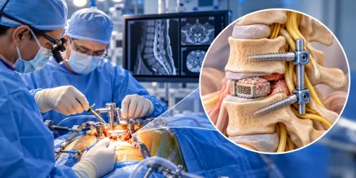
A suspicious spot on a vertebra, disc, epidural space, or nearby soft tissue does not always have one obvious cause. By the end, you will know when imaging and blood tests are enough, when tissue is needed, how the sample is collected and tested, and how to choose a safe diagnostic pathway in Jaipur.
Key takeaways
- Biopsy distinguishes infection, cancer, fracture, and degenerative tissue changes.
- Imaging and blood tests can answer some cases without tissue sampling.
- CT and MRI help plan the safest minimally invasive biopsy route.
- Bring scans, reports, medication details, and previous treatment records.
What a spinal lesion biopsy is designed to distinguish
A spinal lesion biopsy is designed to identify what abnormal tissue is, not merely confirm that MRI found it. Imaging can show destruction, swelling, or a mass, but it often cannot reliably separate infection from cancer, a benign fracture, or degenerative change. Tissue may be needed before chemotherapy, radiotherapy, or major spine surgery.
| Condition | What biopsy can confirm or separate | Why imaging may not settle it |
|---|---|---|
| Infection | Bacterial, tuberculosis, or fungal infection; pus or infected bone | Infection can resemble a tumour or inflammatory lesion |
| Metastatic cancer | Cancer spread from another organ | Metastases can mimic myeloma, lymphoma, or infection |
| Primary bone tumour | A tumour arising in the vertebra, including benign and malignant types | Different tumours can share the same destructive appearance |
| Myeloma or lymphoma | Marrow-based blood cancer involving the spine | MRI may show replacement of marrow without identifying its cause |
| Inflammatory disease | Inflammatory tissue or granulomatous disease | Infection and inflammation can produce similar enhancement |
| Fracture or benign lesion | Healing fracture, haemangioma, cyst, or other non-cancerous tissue | A benign collapse can resemble tumour-related bone destruction |
A spinal lesion biopsy in Jaipur is a diagnostic pathway, not one standard operation. CT-guided needle sampling commonly targets a vertebral or paraspinal lesion; aspiration collects fluid or pus, while an open biopsy removes tissue surgically.
A negative or nondiagnostic result does not prove the lesion is benign: necrotic tissue, too little material, or poor specimen handling can hide the diagnosis. Persistent concern can require repeat image-guided sampling or open biopsy.
When imaging or blood tests can answer the question without biopsy
A spine biopsy in Jaipur is unnecessary when MRI or CT shows a characteristic, low-risk finding that matches the symptoms. Examples include typical vertebral haemangioma, uncomplicated degenerative disease, or an osteoporotic compression fracture with a clear clinical history.
Blood tests can also settle some questions: a complete blood count, calcium, alkaline phosphatase, ESR, CRP, or serum protein electrophoresis may support infection, metabolic bone disease, or myeloma.
- MRI can answer the question when the lesion has a classic benign pattern, or when disc, nerve, and soft-tissue findings clearly explain the symptoms.
- CT can identify a typical fracture, dense bone island, or characteristic mineralisation without tissue sampling.
- PET-CT can map known cancer and reveal whether other sites are involved, but increased uptake is not proof of cancer; infection and healing fractures can look similar.
- Follow-up MRI or CT is reasonable for a small, non-aggressive lesion when symptoms are stable and the scan shows no cortical destruction, soft-tissue mass, or rapid growth.
Imaging and blood tests leave too much uncertainty when a lesion is solitary, enlarging, destructive, or atypical; when MRI cannot separate infection from tumour; or when treatment would differ between metastasis, primary bone tumour, tuberculosis, and inflammatory disease. Biopsy becomes important when cultures, histology, immunohistochemistry, flow cytometry, or molecular testing will determine treatment.
Discordance between symptoms, laboratory results, and scans is another reason not to rely on follow-up alone.
How the safest biopsy route is planned from MRI and CT
The safest target is the most diagnostically useful solid tissue that can be reached without crossing the spinal cord, nerve roots, major blood vessels, or unstable bone. MRI maps the lesion’s soft-tissue, epidural and marrow components; CT shows bone destruction, calcification and a safe needle corridor.
The radiologist and spine team match the lesion’s location to the access route:
- Vertebral-body lesion: CT-guided needle placement through a transpedicular or posterolateral route, depending on the level and bone anatomy.
- Posterior-element lesion: a short posterior route through the lamina, pedicle, facet or spinous-process region, with CT confirming the bony trajectory.
- Disc or endplate abnormality: sampling of the disc or adjacent bone when infection is suspected, while avoiding the thecal sac and nerve roots.
- Epidural mass: biopsy only when a protected corridor exists; urgent surgical assessment takes priority if the mass is compressing the cord or causing instability.
- Paraspinal soft-tissue mass: CT guidance is common, while ultrasound or fluoroscopy can suit selected superficial or clearly visible targets.
The operator avoids necrotic, haemorrhagic or purely cystic areas unless fluid or pus is the diagnostic specimen. A CT-guided minimally invasive spine biopsy is usually preferred; fluoroscopy suits selected bony targets, while open biopsy is reserved for inaccessible lesions or cases requiring decompression or stabilisation.
The planned route also determines whether bone cores, soft-tissue cores or fluid go to pathology, microbiology, or both.
What happens before, during, and after minimally invasive sampling
Expect a small skin puncture rather than a large incision. Before a minimally invasive spine biopsy, review medicines—especially blood thinners— allergies, pregnancy status, and recent blood tests with the team. If sedation is planned, follow the facility’s fasting instructions and arrange an escort home.
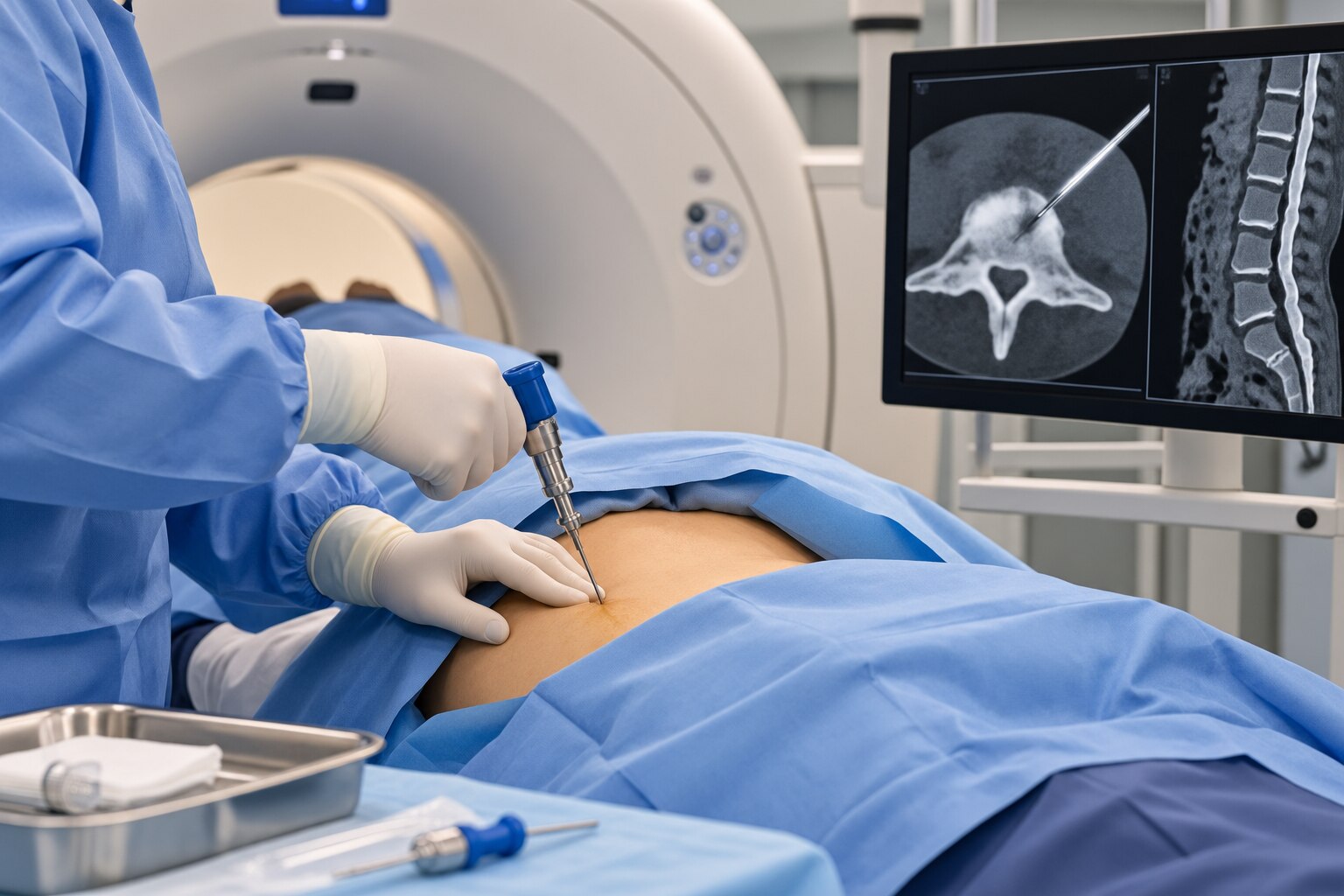
- You will lie in the position selected from your MRI or CT plan. The operator cleans the skin, gives local anaesthetic, and may add intravenous sedation; general anaesthesia is not usually needed.
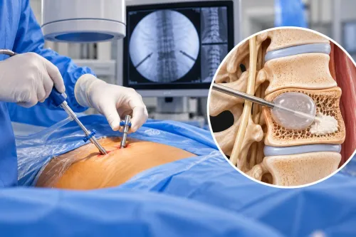
- Using CT or fluoroscopy, the operator advances a biopsy needle along the safest route to the planned bone, disc, epidural, or paraspinal target. You may feel pressure, but report sharp or radiating pain immediately.
- The needle removes one or more small cores or fluid samples. Ask whether separate portions are being sent for histopathology, bacterial, fungal, or tuberculosis testing when infection is possible.
- Staff apply a dressing and observe you during recovery. Sedation generally means no driving, alcohol, or strenuous activity for the period specified by the facility; keep the puncture site clean and follow its lifting instructions.
Mild soreness or bruising can follow. Seek urgent advice for increasing pain, fever, redness, drainage, new weakness or numbness, severe headache, or difficulty urinating. Bleeding, infection, nerve irritation, or a nondiagnostic sample are recognised risks; a negative sample does not prove the lesion is benign if imaging or symptoms remain concerning.
How to prepare for a spine biopsy consultation in Jaipur
Bring the actual MRI or CT images on a disc, portal link, or film, not only the written report. Also bring prior scans for comparison, your medication list, allergies, previous cancer or tuberculosis history, kidney disease, bleeding disorders, and details of blood thinners.
At a spine biopsy consultation in Jaipur, ask these questions:
- Who will perform the sampling, and will CT, fluoroscopy, or another imaging method guide the needle? Confirm that the planned route avoids the spinal cord, nerve roots, major vessels, and unstable bone.
- What exact diagnosis is being tested: infection, cancer, inflammatory disease, or another lesion? Ask whether the specimen will go to histopathology, bacterial culture, fungal culture, and mycobacterial culture.
- How will each portion be preserved? Fresh tissue may be needed for culture, flow cytometry, immunohistochemistry, or molecular testing; placing every fragment in formalin can make culture impossible.
- Which pathology laboratory will receive it, and does the microbiology team know the suspected diagnosis before collection? Confirm the laboratory has an established process for these specimens.
- Will sedation be used, and do you need an escort? Ask when you can drive, work, exercise, and expect the preliminary and final results to be reviewed.
A spine biopsy doctor in Jaipur should also explain whether platelet and clotting tests are needed. Do not stop an anticoagulant without the prescribing clinician’s instruction. Dr Rahul Temani can help coordinate the treating specialist’s question with the operator and laboratory when arranging a spine biopsy in Jaipur.
Related service
Advanced Spine Biopsy Treatment for Accurate Diagnosis Advanced Spine Biopsy Treatment for Accurate Diagnosis Spine Biopsy A spine biopsy is a minimally invasive diagnostic procedure used to collect a small sample of bone or tissue from the spine to detect... View service → |
Frequently asked questions
What is a spinal lesion biopsy designed to distinguish?
It identifies abnormal tissue and helps distinguish infection, cancer, benign fracture, and degenerative change when imaging cannot provide a reliable diagnosis.
When can imaging or blood tests avoid a spine biopsy?
A biopsy may not be needed when MRI, CT, blood tests, clinical findings, or follow-up imaging provide a sufficiently clear diagnosis for treatment.
How are MRI and CT used to plan a spine biopsy?
MRI shows the lesion, nerves, spinal cord, and soft tissues. CT maps the bone and helps the specialist choose a safe needle path that avoids vital structures.
What should you bring to a spine biopsy consultation in Jaipur?
Bring MRI and CT images with reports, blood-test results, medication and allergy lists, previous treatment records, and information about symptoms and their timing.
Keywords