Jaipur
08048035249
+919983718693

Treatment

UBE Spine Surgery (Unilateral Biportal Endoscopy)

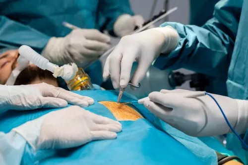
UBE Spine Surgery (Unilateral Biportal Endoscopy) for Minimally Invasive Spine Treatment Unilateral Biportal Endoscopy, commonly known as UBE Spine Surgery, is an advanced minimally invasive procedure used to treat a wide range of spinal disorders, particularly conditions affecting the lumbar spine. This modern surgical technique is transforming spine care by offering precise nerve decompression, minimal tissue damage, and faster recovery compared to traditional open spine surgery. Unlike conventional surgery, UBE uses two small portals — one for a high-definition endoscopic camera and the other for specialized surgical instruments. This dual-portal approach allows the surgeon to directly visualize the spinal structures and perform accurate treatment with minimal disruption to surrounding muscles and tissues. What is UBE Spine Surgery? UBE Spine Surgery is a minimally invasive endoscopic spine procedure that enables surgeons to treat spinal conditions through tiny incisions using real-time camera guidance. The technique provides excellent visualization of the spinal nerves, discs, and surrounding structures while preserving normal anatomy. The primary goals of UBE surgery are to: Relieve nerve compression Remove herniated disc material Decompress the spinal canal Reduce pain and neurological symptoms Preserve muscles and spinal stability Promote faster recovery and rehabilitation Conditions Treated with UBE Spine Surgery UBE is highly effective in treating various spinal disorders, including: Lumbar Disc Herniation Lumbar Canal Stenosis Foraminal Stenosis Degenerative Disc Disease Facet Joint Cysts Sciatica Ligamentum Flavum Hypertrophy Recurrent Disc Herniation Selected Spinal Infections Mild Spinal Instability in selected patients How UBE Spine Surgery is Performed 1. Anesthesia The procedure is usually performed under general or spinal anesthesia. 2. Two Small Portals Two tiny incisions are made near the affected spinal segment: One portal is used for the endoscopic camera The second portal is used for surgical instruments 3. Endoscopic Visualization A high-definition camera provides a magnified and clear view of the spinal structures. 4. Decompression and Treatment The surgeon removes herniated disc material, thickened ligaments, or bone causing nerve compression while preserving healthy tissues. 5. Closure The small incisions are closed with minimal sutures or adhesive strips, resulting in very little scarring. Key Benefits of UBE Spine Surgery Minimally Invasive Technique Small skin incisions Minimal muscle and tissue damage Faster Recovery Early mobilization after surgery Quicker return to daily activities Less Blood Loss Cleaner and safer surgical field Reduced risk of complications Reduced Post-Operative Pain Less need for strong pain medications Improved patient comfort Better Cosmetic Results Minimal scarring due to tiny incisions High-Definition Visualization Precise identification and treatment of nerve compression Shorter Hospital Stay Many patients can be discharged on the same day or within 24 hours Suitable for Elderly Patients Lower surgical stress and faster rehabilitation Recovery After UBE Spine Surgery Recovery following UBE surgery is generally rapid compared to traditional spine surgery. Most patients are able to walk within hours after the procedure and resume light activities within a few days. Physiotherapy and guided rehabilitation may help improve long-term outcomes and spinal strength.

Minimally Invasive Spine Surgery

Minimally Invasive Spine Surgery (MISS) for Faster Recovery & Lasting Pain Relief Minimally Invasive Spine Surgery, commonly known as MISS, is an advanced surgical approach used to treat spinal disorders with minimal disruption to muscles, tissues, and bones. Unlike traditional open spine surgery, MISS uses small incisions, specialized instruments, and real-time imaging guidance to perform precise spinal procedures with reduced tissue damage. This modern technique offers effective treatment for conditions such as herniated discs, spinal stenosis, spinal fractures, spondylolisthesis, and degenerative disc disease while promoting faster recovery and less postoperative discomfort. What is Minimally Invasive Spine Surgery? MISS involves accessing the spine through very small incisions using tubular retractors, endoscopes, microscopes, and image-guided navigation systems. These technologies allow surgeons to reach the affected area accurately without extensive muscle cutting or tissue dissection. The primary goals of MISS are to: Relieve pressure on spinal nerves Stabilize the spine when needed Reduce pain and neurological symptoms Preserve normal spinal anatomy Accelerate recovery and rehabilitation Conditions Treated with MISS Minimally Invasive Spine Surgery is commonly used to treat: Herniated or Slipped Discs Spinal Stenosis (narrowing of the spinal canal) Sciatica and Radiculopathy Spondylolisthesis (slipped vertebra) Spinal Fractures Degenerative Disc Disease Spinal Tumors Spinal Infections Failed Back Surgery Syndrome Common MISS Procedures 1. Microdiscectomy A minimally invasive procedure used to remove herniated disc material pressing on spinal nerves. 2. Minimally Invasive Laminectomy Removal of a small portion of vertebral bone to relieve nerve compression caused by spinal stenosis. 3. Minimally Invasive Spinal Fusion Stabilization of spinal segments using screws, rods, cages, and bone grafts through small incisions. 4. Vertebroplasty and Kyphoplasty Procedures used to treat compression fractures by injecting bone cement into weakened vertebrae. 5. Endoscopic Spine Surgery An ultra-minimally invasive technique performed using a tiny camera and specialized instruments for precise treatment with minimal tissue trauma. How MISS is Performed 1. Anesthesia The surgery is performed under general or spinal anesthesia depending on the procedure. 2. Small Incisions Tiny incisions are made near the affected spinal area. 3. Image-Guided Access Real-time imaging such as fluoroscopy or navigation systems helps guide the surgeon accurately. 4. Surgical Treatment Specialized instruments are used to decompress nerves, remove damaged tissue, or stabilize the spine. 5. Closure The small incisions are closed with minimal sutures or surgical adhesive. Benefits of Minimally Invasive Spine Surgery Smaller Incisions Usually less than one inch Minimal scarring and better cosmetic results Less Pain and Blood Loss Reduced muscle and tissue damage Less postoperative discomfort Faster Recovery Earlier mobilization and rehabilitation Quicker return to work and daily activities Lower Risk of Infection Smaller wounds reduce the chances of complications Shorter Hospital Stay Many procedures are performed as day-care surgeries or require only one-night admission Preservation of Normal Anatomy Maintains spinal stability and reduces the risk of future problems Recovery After MISS Recovery after MISS is generally much faster compared to traditional open spine surgery. Most patients begin walking within hours or the next day after surgery. Depending on the procedure, many individuals return to light activities within days to weeks. Physical therapy may be recommended to improve strength and flexibility.

Spine Fracture Fixation Surgery For Spinal Stability

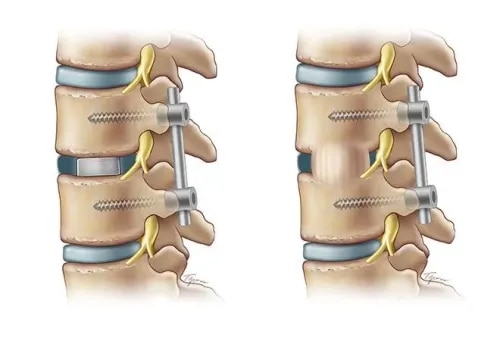
Spine Fracture Fixation Surgery for Spinal Stability & Pain Relief Spine Fracture Fixation is an advanced surgical procedure performed to stabilize broken or collapsed vertebrae, restore spinal alignment, protect the spinal cord, and promote proper healing. Spinal fractures can be painful, disabling, and potentially life-threatening if left untreated. These fractures may occur due to trauma, osteoporosis, tumors, or sports-related injuries. While minor fractures may heal with rest and bracing, severe or unstable fractures often require surgical fixation to restore stability and prevent long-term complications. What is a Spine Fracture? A spinal fracture occurs when one or more vertebrae in the spinal column break due to excessive force or weakened bone structure. Depending on the severity, these fractures can lead to spinal instability, deformity, nerve compression, chronic pain, or even paralysis if not managed properly. Common Causes of Spinal Fractures Road traffic accidents and high-impact trauma Falls, especially in elderly individuals Sports-related injuries Osteoporosis-related compression fractures Tumors or metastatic cancers weakening the bones Violent injuries such as gunshot wounds What is Spine Fracture Fixation? Spine Fracture Fixation is a surgical technique that uses specialized implants such as screws, rods, plates, cages, or bone cement to stabilize the fractured spine. The procedure aims to: Realign fractured vertebrae Stabilize the spinal column Relieve pressure on spinal nerves if needed Promote proper bone healing Prevent spinal cord injury and deformity Depending on the patient’s condition and fracture type, the surgery may be performed using open surgery or minimally invasive spine surgery techniques. Types of Spine Fracture Fixation Procedures 1. Pedicle Screw Fixation Specialized screws are inserted into the vertebrae and connected with rods to stabilize the fractured spinal segment and restore alignment. 2. Vertebroplasty and Kyphoplasty Bone cement is injected into a collapsed vertebra to strengthen it, reduce pain, and restore vertebral height. These procedures are commonly used for osteoporotic compression fractures. 3. Corpectomy and Fusion The damaged vertebra is removed and replaced with an implant or cage, followed by spinal fusion using bone grafts and instrumentation. 4. Spinal Fusion Surgery Two or more vertebrae are permanently joined together to eliminate abnormal motion and provide long-term stability. How Spine Fracture Fixation is Performed 1. Anesthesia The procedure is performed under general anesthesia. 2. Surgical Access A small or standard incision is made depending on whether minimally invasive or open surgery is planned. 3. Fracture Stabilization The fractured vertebrae are realigned and stabilized using screws, rods, plates, cages, or bone cement. 4. Nerve Decompression If nerves or the spinal cord are compressed, decompression is performed to relieve pressure. 5. Fusion and Closure Bone grafts may be placed to encourage spinal fusion, and the incision is closed with sutures or staples. Benefits of Spine Fracture Fixation Immediate stabilization of the spine Significant pain relief Faster recovery and rehabilitation Restoration of spinal alignment and vertebral height Reduced risk of spinal cord or nerve damage Improved mobility and quality of life Prevention of long-term deformity and disability Recovery After Spine Fracture Fixation Recovery time depends on the severity of the fracture and the type of surgery performed. Most patients begin assisted movement within a few days after surgery. Physical therapy and rehabilitation are essential for restoring strength, flexibility, and mobility. Complete healing and fusion may take several months.

Laminectomy Surgery for Spinal Stenosis & Nerve Compression

Laminectomy Surgery for Spinal Nerve Compression & Pain Relief Laminectomy is a common spinal surgery performed to relieve pressure on the spinal cord or nerves caused by conditions such as spinal stenosis, herniated discs, bone spurs, or spinal tumors. The procedure involves removing the lamina — the back portion of a vertebra that covers the spinal canal — to create more space and reduce nerve compression. This surgical treatment is highly effective in reducing chronic back pain, leg pain, numbness, and weakness while improving mobility and overall quality of life. What is the Lamina? The lamina is a small bony arch located at the back of each vertebra. It forms the protective covering of the spinal canal and helps shield the spinal cord and nerves. However, age-related degeneration, enlarged ligaments, bone spurs, or damaged discs can narrow the spinal canal and compress nearby nerves, leading to pain and neurological symptoms. When is Laminectomy Needed? Laminectomy is usually recommended when conservative treatments such as medications, physiotherapy, lifestyle changes, or spinal injections fail to provide adequate relief. It may be advised for patients experiencing: Chronic back or neck pain Leg or arm pain (sciatica or radiculopathy) Tingling, numbness, or weakness in the limbs Difficulty walking or maintaining balance Loss of bladder or bowel control in severe cases Common Conditions Treated with Laminectomy Lumbar Spinal Stenosis Cervical or Thoracic Spinal Stenosis Herniated Disc Spinal Tumors Spondylolisthesis Osteoarthritis-related Bone Spurs How is Laminectomy Performed? 1. Anesthesia The surgery is performed under general or spinal anesthesia to ensure patient comfort. 2. Incision A small incision is made over the affected area of the spine. 3. Surgical Exposure The surrounding muscles are gently moved aside to provide access to the spinal column. 4. Lamina Removal The surgeon carefully removes part or all of the lamina to relieve pressure on the spinal cord and nerves. 5. Optional Spinal Fusion In some patients, spinal fusion or stabilization may be performed along with laminectomy to improve spinal stability. 6. Closure The incision is closed using sutures or staples and covered with a sterile dressing. Benefits of Laminectomy Surgery Effective relief from nerve-related pain Reduced numbness, tingling, and muscle weakness Improved walking ability and mobility Better spinal nerve decompression Enhanced quality of life and daily functioning Faster return to normal activities after recovery Recovery After Laminectomy Most patients begin walking within a day after surgery. Recovery time depends on the extent of the procedure and the patient’s overall health. Light daily activities can usually be resumed within a few weeks, while complete recovery may take several months. Physical therapy is often recommended to improve strength and flexibility.

ACDF (Anterior Cervical Discectomy and Fusion): Modern Neck Spine Surgery

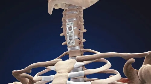
ACDF Surgery (Anterior Cervical Discectomy and Fusion) Anterior Cervical Discectomy and Fusion, commonly known as ACDF, is a highly effective spinal surgery used to treat various cervical spine conditions such as herniated discs, degenerative disc disease, cervical spondylosis, and spinal stenosis. The procedure involves removing a damaged disc through the front of the neck (anterior approach) and fusing the adjacent vertebrae to restore spinal stability, relieve nerve compression, and reduce pain. ACDF is one of the most commonly performed cervical spine surgeries and is known for its excellent outcomes in relieving neck pain, arm pain, numbness, and weakness caused by nerve compression. What is ACDF? ACDF is a specialized cervical spine procedure designed to: Remove a damaged or herniated disc Relieve pressure on the spinal cord and nerve roots Restore normal spinal alignment Stabilize the cervical spine by fusing vertebrae together using a bone graft, cage, and metal implants The surgery helps eliminate painful motion between vertebrae while improving overall neck stability and function. When is ACDF Recommended? ACDF surgery is generally recommended when conservative treatments such as medications, physiotherapy, injections, or rest fail to provide adequate relief. It is particularly beneficial for patients experiencing: Persistent neck pain Radiating arm pain (cervical radiculopathy) Numbness or tingling in the arms or hands Weakness in the upper limbs Spinal cord compression (cervical myelopathy) Herniated cervical discs Degenerative disc disease Cervical spondylosis Spinal instability or trauma How ACDF Surgery is Performed 1. Anesthesia The procedure is performed under general anesthesia to ensure patient comfort during surgery. 2. Small Neck Incision A small incision is made in the front of the neck to access the cervical spine. 3. Disc Removal The damaged disc, along with any bone spurs compressing the nerves, is carefully removed. 4. Nerve Decompression Pressure on the spinal cord and nerve roots is relieved to reduce pain and neurological symptoms. 5. Fusion Procedure A bone graft or interbody cage is inserted into the empty disc space to maintain proper spacing and promote fusion. 6. Stabilization A metal plate and screws are placed to stabilize the vertebrae and support healing. Benefits of ACDF Surgery Immediate relief from nerve compression symptoms Effective reduction in neck and arm pain Restores spinal alignment and stability Prevents painful motion between vertebrae Small incision with minimal visible scarring Faster recovery and shorter hospital stay High success rate for cervical radiculopathy treatment Improved mobility and quality of life Recovery After ACDF Surgery Most patients can walk within hours after surgery and are discharged within one or two days, depending on the complexity of the procedure. Mild discomfort during recovery is common and gradually improves over time. Physical therapy and activity modification may be recommended to support healing and restore strength. Complete fusion may take several months, but many patients experience significant symptom relief shortly after surgery.

Advanced Endoscopic Spine Surgery for Back & Neck Pain Relief

Endoscopic Spine Surgery – Advanced Minimally Invasive Treatment for Spine Disorders Endoscopic Spine Surgery is a modern, minimally invasive procedure designed to treat a wide range of spinal conditions through a very small incision using a high-definition endoscope (camera). This advanced technique serves as an effective alternative to traditional open spine surgery by reducing tissue damage, postoperative pain, and recovery time. Endoscopic Spine Surgery is especially beneficial for patients suffering from herniated discs, spinal stenosis, chronic back pain, and sciatica. By providing precise access to the affected spinal area, surgeons can accurately remove the source of nerve compression while preserving healthy muscles and surrounding tissues. Conditions Treated with Endoscopic Spine Surgery Endoscopic Spine Surgery is commonly recommended for: Herniated (slipped) disc Lumbar or cervical disc prolapse Sciatica and leg pain caused by nerve compression Spinal stenosis (narrowing of the spinal canal) Foraminal stenosis Facet joint cysts Failed Back Surgery Syndrome Recurrent disc herniation Degenerative disc disease Types of Endoscopic Spine Surgeries Transforaminal Endoscopic Discectomy (TFED) A minimally invasive procedure used to treat disc herniation causing leg pain and sciatica through a transforaminal approach. Interlaminar Endoscopic Discectomy (ILED) Ideal for centrally located disc herniations, allowing direct access to the affected spinal canal. Endoscopic Decompression Performed to relieve pressure on nerves caused by spinal canal stenosis or foraminal narrowing. Cervical Endoscopic Surgery Used for treating disc problems and nerve compression in the neck (cervical spine) region. Benefits of Endoscopic Spine Surgery Minimally Invasive Technique Tiny incision with minimal scarring Reduced muscle and tissue damage Lower risk of infection and complications Faster Recovery Shorter hospital stay, often same-day discharge Quicker return to work and daily activities Faster rehabilitation compared to open surgery Reduced Pain Less postoperative discomfort Reduced dependency on pain medications Minimal Blood Loss Safer for elderly patients and individuals with medical comorbidities High Surgical Precision High-definition visualization of the spine Accurate removal of damaged disc material and compressed tissue Better Cosmetic Outcome Small scars that are barely visible Local or Mild Sedation In selected cases, surgery can be performed under local anesthesia or mild sedation, reducing anesthesia-related risks Recovery After Endoscopic Spine Surgery Most patients are encouraged to walk within a few hours after surgery. Because the procedure causes minimal tissue disruption, recovery is significantly faster than traditional open spine surgery. Patients can often resume light activities within days, although complete healing depends on the severity of the spinal condition and overall health.

Advanced Spinal Deformity Correction Surgery In Jaipur

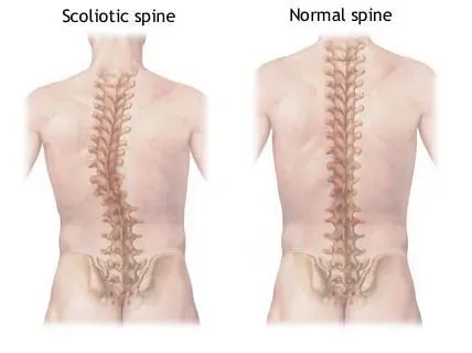
Spinal Deformity Correction Surgery Spinal Deformity Correction is a specialized surgical approach designed to treat abnormal curvatures and misalignments of the spine. These deformities can affect posture, mobility, balance, and overall quality of life, often leading to chronic pain, nerve compression, or breathing difficulties if left untreated. Spinal deformities may be congenital, develop during childhood or adolescence, or occur later in life due to degeneration, trauma, osteoporosis, or neurological disorders. Treatment is carefully personalized to restore spinal alignment, relieve symptoms, and improve daily function. What is Spinal Deformity? Spinal deformities refer to abnormal curvatures or structural imbalances of the spine that differ from the normal spinal alignment. Common types include: Scoliosis – Sideways curvature of the spine Kyphosis – Excessive forward rounding or hunchback appearance Lordosis – Increased inward curvature of the lower back Flat Back Syndrome – Loss of the normal lumbar curvature Post-traumatic or degenerative spinal deformities Causes of Spinal Deformity Spinal deformities can develop due to several underlying conditions, including: Congenital spine malformations Adolescent idiopathic scoliosis Osteoporosis and spinal fractures Degenerative disc disease Ankylosing Spondylitis Failed back surgery syndrome Spinal tumors or infections Neuromuscular disorders such as cerebral palsy Treatment Options for Spinal Deformity 1. Non-Surgical Treatments Mild to moderate deformities may often be managed without surgery through: Bracing, especially in adolescents with scoliosis Physiotherapy and posture correction exercises Pain management with medications Chiropractic or manual therapy (in selected cases) Lifestyle modifications and rehabilitation 2. Surgical Treatments Surgery is generally recommended for severe, progressive, or symptomatic spinal deformities. Spinal Fusion Surgery Two or more vertebrae are fused together using rods, screws, and bone grafts to stabilize and straighten the spine. Osteotomy A surgical procedure in which bone is cut and reshaped to restore proper spinal alignment. Vertebral Column Resection (VCR) Part of a vertebra is removed to correct severe or rigid spinal deformities. Expandable Rod Systems Special growing rod systems are used in children to allow continued spinal growth while correcting deformity. Minimally Invasive Spine Surgery Selected deformities may be treated using minimally invasive techniques that involve smaller incisions, reduced muscle damage, and faster recovery. Goals and Benefits of Spinal Deformity Correction Restore proper spinal alignment and balance Relieve chronic back pain and nerve compression Improve posture, mobility, and flexibility Enhance lung function in severe spinal curvature cases Prevent progression of spinal deformity Improve physical activity and independence Boost confidence and body image Recovery After Surgery Recovery depends on the severity of the deformity and the type of surgery performed. Most patients undergo rehabilitation and physical therapy to regain strength and mobility. Modern surgical techniques and advanced spinal instrumentation have significantly improved recovery times and long-term outcomes.

Microdiscectomy Surgery for Slip Disc & Sciatica Relief

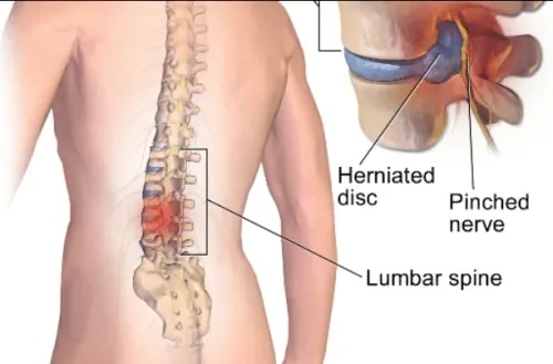
Microdiscectomy Surgery for Herniated Disc & Sciatica Relief Microdiscectomy is a minimally invasive spinal surgery performed to relieve pressure on a spinal nerve caused by a herniated (slipped) disc in the lower back (lumbar spine) or neck (cervical spine). It is one of the most effective procedures for treating sciatica, leg pain, numbness, and nerve compression when non-surgical treatments fail to provide relief. What is a Herniated Disc? The spine is made up of vertebrae separated by soft discs that act as shock absorbers. A herniated disc occurs when the inner gel-like material of the disc pushes through the outer layer and compresses nearby spinal nerves. This pressure can lead to severe pain, weakness, tingling, and difficulty with movement. Symptoms That May Require Microdiscectomy Microdiscectomy may be recommended for patients experiencing: Persistent leg pain (sciatica) Numbness or tingling in the leg or foot Weakness in lower limb muscles Difficulty standing, walking, or sitting for long periods Severe nerve compression symptoms Lack of improvement after 6–8 weeks of medications or physiotherapy What is Microdiscectomy Surgery? Microdiscectomy is a targeted surgical procedure that removes the portion of the herniated disc pressing on the spinal nerve. The surgery is performed using specialized microscopic instruments through a very small incision, allowing minimal disruption to surrounding muscles and tissues. Compared to traditional open spine surgery, microdiscectomy offers faster recovery, reduced pain, and lower risk of complications. Procedure Overview 1. Anesthesia The procedure is usually performed under general or spinal anesthesia. 2. Small Incision A small incision, typically around 1–1.5 inches, is made over the affected spinal area. 3. Muscle Retraction The surrounding muscles are gently moved aside rather than cut, helping preserve normal tissue. 4. Microscope-Assisted Surgery A surgical microscope provides a magnified and precise view of the compressed nerve and disc. 5. Disc Fragment Removal The surgeon carefully removes the herniated portion of the disc that is pressing on the nerve root. 6. Closure & Recovery The incision is closed with minimal sutures, and the patient is moved to recovery. Benefits of Microdiscectomy Surgery Minimally invasive with smaller incision Less muscle and tissue damage Faster recovery and early mobility Short hospital stay or same-day discharge High success rate in relieving sciatica and leg pain Reduced post-operative discomfort Smaller scar and lower infection risk Quicker return to daily activities and work Recovery After Microdiscectomy Most patients begin walking within hours after surgery and return home the same day or the next day. Light activities can usually be resumed within a few weeks, while complete recovery depends on the individual’s condition and rehabilitation process. Following post-surgical exercises and spine care guidelines is essential for long-term success.

TLIF Treatment for Chronic Lower Back Pain

TLIF Surgery (Transforaminal Lumbar Interbody Fusion) Transforaminal Lumbar Interbody Fusion, commonly known as TLIF, is an advanced spinal fusion procedure used to treat chronic lower back pain caused by conditions such as disc degeneration, spinal instability, or spondylolisthesis. The surgery involves removing a damaged spinal disc and fusing two vertebrae together using a bone graft and specialized implants to restore long-term spinal stability. This minimally invasive technique is highly effective in relieving sciatica, nerve compression, and instability in the lumbar spine while promoting faster recovery and reduced tissue damage. What is TLIF? TLIF is a surgical technique in which the surgeon approaches the spine from the side of the lower back through a transforaminal route. During the procedure, the damaged disc is removed, compressed nerves are relieved, and a bone graft or interbody cage is placed into the disc space. Screws and rods are then used to stabilize the spine and support proper fusion between the vertebrae. When is TLIF Recommended? TLIF surgery is generally recommended when non-surgical treatments such as medications, physiotherapy, injections, or lifestyle modifications fail to provide lasting relief. It is commonly advised for patients suffering from: Degenerative Disc Disease Spondylolisthesis (slipped vertebra) Recurrent Disc Herniation Spinal Instability Foraminal Stenosis Failed Back Surgery Syndrome Chronic Sciatica How TLIF Surgery is Performed 1. Anesthesia The procedure is performed under general anesthesia to ensure patient comfort throughout the surgery. 2. Small Incision A small incision is made near the midline of the lower back. 3. Surgical Access The surgeon gently retracts the surrounding muscles and accesses the spine through a transforaminal approach. 4. Disc Removal The damaged or herniated disc is carefully removed to relieve pressure on the spinal nerves. 5. Bone Graft or Cage Placement A spacer or cage filled with bone graft material is inserted between the vertebrae to maintain disc height and encourage fusion. 6. Stabilization Specialized screws and rods are placed to stabilize the spinal segment and maintain proper alignment. 7. Fusion Process Over time, the bone graft helps the adjacent vertebrae fuse into a single solid bone, providing long-term stability. Benefits of TLIF Surgery Minimally invasive procedure with smaller incisions Effective decompression of spinal nerves Improved spinal stability and alignment Long-lasting relief from back and leg pain Reduced blood loss and muscle damage Shorter hospital stay and quicker recovery Lower risk of postoperative instability compared to traditional approaches Recovery After TLIF Surgery Most patients are encouraged to walk within a day after surgery. Recovery time varies depending on the patient’s overall health and the complexity of the procedure. Light activities may resume within a few weeks, while complete spinal fusion can take several months. Physical therapy and proper rehabilitation play an important role in achieving the best outcomes.

Advanced Spine Biopsy Treatment for Accurate Diagnosis

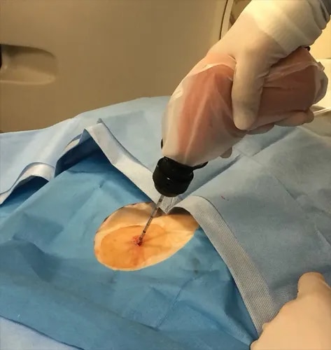
Spine Biopsy A spine biopsy is a minimally invasive diagnostic procedure used to collect a small sample of bone or tissue from the spine to detect infections, tumors, inflammation, or other spinal abnormalities. It plays a vital role in identifying the exact cause of spinal lesions or persistent back pain, especially when MRI or CT scan findings are unclear. What is a Spine Biopsy? During a spine biopsy, a small tissue or bone sample is removed using a needle or a minor surgical procedure. The collected sample is examined in a pathology laboratory to determine whether the condition is caused by cancer, infection, tuberculosis, or other spinal diseases. Spine biopsies may be performed in the cervical (neck), thoracic (mid-back), or lumbar (lower back) spine depending on the location of the abnormality. Types of Spine Biopsy Needle Biopsy (Percutaneous Biopsy) Minimally invasive procedure Performed under CT scan or fluoroscopy guidance Usually done under local anesthesia Faster recovery with minimal discomfort Open Biopsy (Surgical Biopsy) Performed in an operating room under general anesthesia Recommended when a larger tissue sample is required Used when needle biopsy is not feasible Why is Spine Biopsy Performed? A spine biopsy helps doctors: Diagnose spinal tumors (benign or malignant) Detect spinal infections such as tuberculosis or osteomyelitis Investigate unexplained back pain with abnormal imaging findings Confirm metastatic cancer involving the spine Plan the most effective treatment strategy Indications for Spine Biopsy Abnormal MRI or CT scan findings Suspected bone cancer or spinal metastasis Suspected spinal tuberculosis or infection Persistent back pain not improving with treatment Vertebral collapse or unexplained spinal fracture How the Procedure is Done The patient is positioned in a CT or fluoroscopy suite The biopsy area is sterilized and numbed with anesthesia A biopsy needle is carefully guided into the affected spinal area Tissue or bone samples are collected for laboratory analysis The procedure typically takes 30–60 minutes Benefits of Spine Biopsy Provides accurate diagnosis of spinal conditions Minimally invasive and safe procedure Short recovery time Helps avoid unnecessary treatments or surgery Assists doctors in planning targeted treatment options

Kyphoplasty Treatment for Spine Fractures in Jaipur

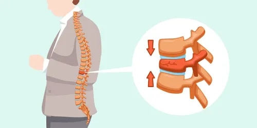
Kyphoplasty Treatment for Spine Fractures in Jaipur Kyphoplasty is an advanced, minimally invasive spine procedure used to treat vertebral compression fractures (VCFs) caused by osteoporosis, trauma, or spinal tumors. This modern technique is highly effective in relieving severe back pain, restoring spinal stability, and improving overall quality of life. For patients suffering from sudden or chronic back pain due to spinal fractures, kyphoplasty offers rapid pain relief, quicker recovery, and long-lasting results. Performed by experienced spine specialists, this procedure has become a preferred treatment option for fragile spine conditions, especially in elderly patients. What Is a Vertebral Compression Fracture (VCF)? A vertebral compression fracture occurs when one of the bones in the spine (vertebra) collapses or becomes compressed. This condition is most commonly seen in individuals with osteoporosis, but it can also result from trauma, injury, or cancer affecting the spine. Common Causes of VCF Osteoporosis (weak and brittle bones) Spinal trauma or accidents Metastatic cancer or spinal tumors Long-term steroid use Age-related bone degeneration Symptoms of Vertebral Compression Fracture Sudden or severe back pain Pain that worsens with movement or standing Loss of height over time Stooped or hunched posture (kyphosis) Reduced mobility and daily activity limitations If left untreated, these fractures can lead to chronic pain, spinal deformity, and decreased quality of life. What Is Kyphoplasty? Kyphoplasty is a minimally invasive spine surgery designed to treat vertebral compression fractures by stabilizing the fractured bone and restoring its height. Step-by-Step Procedure A small incision is made near the affected vertebra A specialized balloon is inserted into the collapsed vertebra The balloon is gently inflated to restore the vertebra’s height and shape The created cavity is filled with medical-grade bone cement The cement hardens quickly, stabilizing the bone The procedure is usually performed under local or general anesthesia and typically takes less than an hour per vertebra. Who Needs Kyphoplasty? Kyphoplasty is recommended for patients who: Have painful vertebral compression fractures Do not respond to medications or conservative treatment Experience severe back pain affecting daily life Have osteoporosis-related spine fractures Show progressive spinal deformity or height loss Early intervention can prevent complications and significantly improve recovery outcomes. Benefits of Kyphoplasty Treatment Kyphoplasty offers multiple advantages compared to traditional spine surgery: 1. Rapid Pain Relief Most patients experience significant pain reduction within 24–48 hours after the procedure. 2. Minimally Invasive Technique Small incision, minimal tissue damage, and lower risk of complications. 3. Short Recovery Time Patients can usually resume normal activities within a few days. 4. Restoration of Vertebral Height Helps correct spinal deformity and improves posture. 5. Stabilization of Fracture Prevents further collapse of the affected vertebra. 6. Improved Mobility and Function Allows patients to return to daily activities quickly. 7. Reduced Dependence on Pain Medication Decreases the need for long-term painkillers. Is Kyphoplasty Safe? Yes, kyphoplasty is considered a safe and effective procedure when performed by an experienced spine surgeon. The use of advanced imaging techniques ensures precise placement of instruments and bone cement. Low Risk Factors Include: Minimal bleeding Low infection risk Quick hospital discharge (often same-day or next-day) Recovery After Kyphoplasty Recovery after kyphoplasty is usually quick and smooth. Post-Procedure Expectations Immediate or early pain relief Walking within a few hours after the procedure Return to daily activities within a few days Follow-up care and monitoring Rehabilitation Tips Light physical activity as advised Avoid heavy lifting for a few weeks Maintain good posture Start physiotherapy if recommended Why Choose Expert Spine Care for Kyphoplasty? Choosing an experienced spine specialist ensures: Accurate diagnosis and case selection Safe and precise surgical technique Better outcomes and faster recovery Comprehensive pre- and post-treatment care Advanced spine centers use state-of-the-art technology and minimally invasive techniques to deliver the best results. Kyphoplasty vs. Conservative Treatment While mild fractures may heal with rest, medications, and bracing, kyphoplasty is preferred when: Pain is severe and persistent Mobility is significantly reduced There is risk of spinal deformity Conservative treatment fails Kyphoplasty provides faster and more effective relief compared to prolonged non-surgical care. Conclusion Kyphoplasty is a highly effective, minimally invasive solution for treating vertebral compression fractures. By stabilizing the spine, relieving pain, and restoring vertebral height, it helps patients regain independence and improve their overall quality of life. If you are experiencing persistent back pain due to osteoporosis or spine injury, consulting a spine specialist for kyphoplasty treatment can help you return to a pain-free and active lifestyle. Kyphoplasty in Jaipur Best Kyphoplasty Surgeon in Jaipur Spine Fracture Treatment Jaipur Vertebral Compression Fracture Treatment Osteoporosis Spine Fracture Treatment Minimally Invasive Spine Surgery Jaipur Back Pain Treatment Jaipur Spine Specialist in Jaipur Balloon Kyphoplasty Procedure Spine Surgery for Fracture Jaipur

Kyphosis, Scoliosis & Occipito-Cervical Fusion Surgery

Kyphosis, Scoliosis & Occipito-Cervical Fusion Surgery Spinal deformities such as kyphosis and scoliosis can significantly affect posture, balance, mobility, and overall quality of life. In advanced or complex cases—especially when the upper cervical spine is unstable—specialized surgical procedures like occipito-cervical fusion may be required to restore spinal stability and protect critical neurological structures such as the spinal cord and brainstem. With advancements in spine surgery, these conditions can now be effectively managed through a combination of non-surgical treatments and minimally invasive or corrective surgical techniques, helping patients return to a healthier, pain-free life. What is Kyphosis? (Hunchback Spine Deformity) Kyphosis is a spinal condition characterized by an excessive forward curvature of the upper back, leading to a rounded or “hunchback” appearance. While mild kyphosis may not cause significant problems, severe cases can lead to pain, deformity, and breathing issues. Common Causes of Kyphosis Osteoporosis (weakening of bones) Scheuermann’s Disease (developmental disorder in adolescents) Degenerative Disc Disease Spinal Trauma or Fractures Congenital Spine Deformities (present at birth) Symptoms of Kyphosis Noticeable rounding of the upper back Persistent back pain or stiffness Fatigue due to muscle strain Reduced mobility and flexibility Difficulty breathing in severe deformities Early diagnosis and treatment are essential to prevent progression and complications. What is Scoliosis? (Sideways Spine Curvature) Scoliosis is a condition in which the spine curves abnormally to the side, often forming an “S” or “C” shape. It commonly develops during adolescence but can also occur in adults due to aging and degenerative changes in the spine. Common Symptoms of Scoliosis Uneven shoulders, waist, or hips Visible curvature of the spine Back pain or discomfort Muscle imbalance or fatigue Reduced flexibility and restricted movement Breathing problems in severe cases Scoliosis can range from mild to severe and may worsen over time if left untreated. When is Spine Surgery Required? While many cases of kyphosis and scoliosis can be managed with non-surgical treatments such as physiotherapy, bracing, and medications, surgery may be necessary in certain situations. Indications for Spine Surgery Severe or progressive spinal curvature Failure of conservative (non-surgical) treatments Chronic pain affecting daily activities Significant cosmetic deformity Neurological symptoms (numbness, weakness, nerve compression) Spinal cord compression or risk of permanent damage Timely surgical intervention can prevent further complications and improve long-term outcomes. What is Occipito-Cervical Fusion? Occipito-cervical fusion is an advanced spine surgery performed to stabilize the junction between the skull (occiput) and the upper cervical spine (neck region). This procedure is typically recommended for patients with instability, deformity, trauma, tumors, or congenital abnormalities affecting the upper spine. During the procedure, specialized implants such as rods, screws, and bone grafts are used to fuse the skull to the upper cervical vertebrae, creating a stable and secure structure. Conditions Treated with Occipito-Cervical Fusion Upper cervical spine instability Congenital craniovertebral junction anomalies Trauma or fractures of the upper spine Tumors or infections affecting the cervical spine Severe deformities causing neurological compromise Rheumatoid arthritis affecting cervical joints Benefits of Occipito-Cervical Fusion Surgery This highly specialized procedure offers several important benefits: Stabilizes the upper spine and skull junction Protects the spinal cord and brainstem from injury Reduces neurological symptoms such as weakness or numbness Corrects spinal deformity and alignment Prevents further progression of instability Improves overall function and quality of life Recovery and Rehabilitation Recovery after spinal deformity correction or occipito-cervical fusion depends on the severity of the condition and the complexity of the surgery. Post-Surgery Care Includes: Hospital stay for monitoring and recovery Pain management and wound care Gradual mobilization under supervision Physiotherapy and rehabilitation programs Regular follow-up with the spine specialist Most patients experience significant improvement in pain, posture, and neurological function over time. Comprehensive Spine Care Approach Effective treatment of kyphosis, scoliosis, and complex cervical spine conditions involves a multidisciplinary approach: Detailed clinical evaluation and imaging (X-ray, MRI, CT scan) Personalized treatment planning Conservative care (when appropriate) Advanced surgical techniques for correction and stabilization Long-term rehabilitation and lifestyle guidance Kyphosis and scoliosis are serious spinal conditions that can impact both physical health and confidence. In complex cases involving upper spine instability, occipito-cervical fusion surgery provides a safe and effective solution to restore stability, protect neurological structures, and improve overall quality of life. If you or your loved one is experiencing symptoms of spinal deformity or chronic neck and back pain, early consultation with a spine specialist can ensure timely diagnosis and the best possible treatment outcomes.

Subcategory Tags

Phone Number

08048035249

Please keep 0 before dialling the number.

Email Address temsa08@gmail.com

Mon-Thu: 10 AM - 2 PM • Fri: 3 PM - 7AM

Other Website Visit our other website
Address Rajasthan Hospital, 86, opp. RAJASTHAN JAIPURIA HOSPITAL, Kailash Puri Colony, Milap Nagar, Jaipur, Rajasthan 302018, India

Jaipur, India, 302018

footerhc