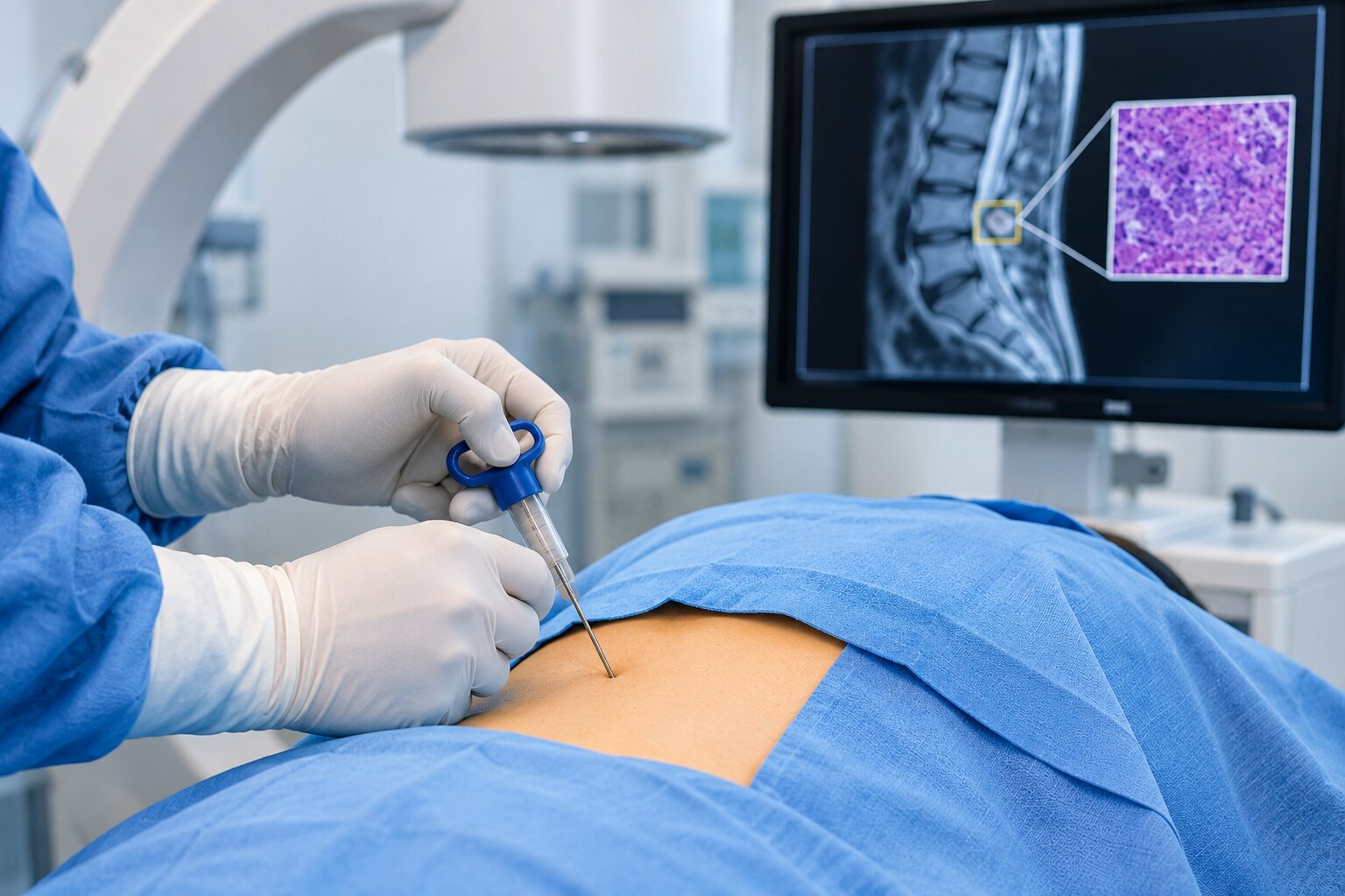
The right biopsy depends on what the scans show, where the abnormality sits, how much tissue the diagnosis requires, and whether a needle can reach it without crossing critical structures. By the end, you will be able to compare biopsy methods, understand route selection, and identify the questions to ask before consenting to a procedure.
Key takeaways
- Confirm a biopsy will change treatment before scheduling it.
- Choose fine-needle, core-needle or open biopsy based on tissue needed.
- Plan the route around nerves, blood vessels and the suspected lesion.
- Use surgery instead when a percutaneous biopsy cannot provide safe, useful tissue.
Start with the lesion, not the biopsy needle
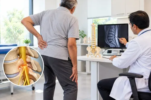
An incidental vertebral lesion does not automatically need a biopsy. Doctors first compare its MRI and CT appearance with symptoms, previous scans, and risks such as cancer or infection. A bone abnormality biopsy becomes important when imaging cannot distinguish a benign change from tumour or infection, or when tissue results will change treatment.
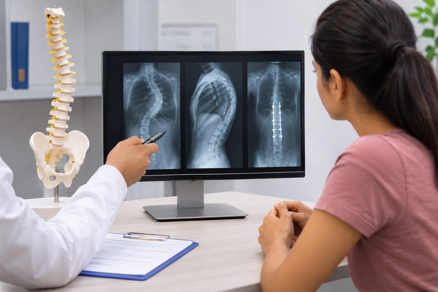
MRI maps marrow replacement, epidural extension, nerve compression and soft-tissue involvement. CT shows cortical destruction, mineralised tumour matrix, pedicle anatomy and the safest needle corridor.
| Imaging | Shows best | Decision value |
|---|---|---|
| MRI | Marrow, epidural space, nerves and soft tissue | Identifies the biologically active target and urgent compression |
| CT | Cortex, mineralisation, pedicles and vessels | Confirms bone destruction and plans the needle trajectory |
| Both | Lesion extent and spinal anatomy | Prevents sampling a nonrepresentative or unsafe area |
For most accessible lesions, CT-guided percutaneous core biopsy is the starting option because it provides tissue architecture. The route must be planned with the surgeon who would perform definitive treatment; a tumour-contaminated needle tract may need removal later.
A transpedicular route suits many thoracic and lumbar vertebral-body lesions when the pedicle is intact and large enough to avoid the canal, nerve roots and vessels. It is a poor choice when the pedicle is destroyed, very small or itself abnormal.
Cervical lesions need separate planning: the carotid and vertebral arteries, airway, oesophagus, spinal cord and nerve roots leave little margin for error, so CT-guided posterior, posterolateral or anterior access depends on cross-sectional imaging.
Fine-needle, core-needle or open biopsy: what tissue is needed?
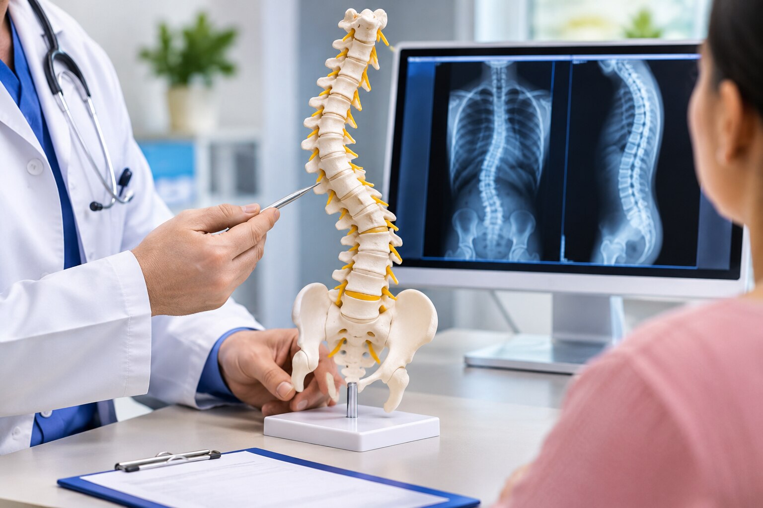
A fine-needle sample shows individual cells; a core sample preserves tissue architecture, which often determines the diagnosis. For most accessible vertebral lesions, CT-guided percutaneous core biopsy is the usual starting choice.
| Option | What it shows | When it applies |
|---|---|---|
| Fine-needle aspiration | Cells for cytology; limited architectural information | A fluid-filled lesion, suspected recurrence, or a case where a rapid cellular assessment is enough |
| Core-needle biopsy | Cylinders of tissue showing architecture, matrix, necrosis and tumour invasion | Most solid bone lesions, including suspected primary tumours or metastases |
| Open surgical biopsy | A larger, deeper specimen from the lesion | An inaccessible target, repeated nondiagnostic needle samples, or a lesion requiring extensive tissue for classification |
Fine-needle aspiration alone can miss primary bone tumours, some lymphomas and infections because the diagnosis depends on how cells are arranged. Lymphoma or suspected metastatic cancer may also need fresh, unfixed tissue for flow cytometry, immunohistochemistry or molecular testing.
If infection is possible, state that on the request before the procedure. Send separate sterile material for:
- Aerobic and anaerobic bacterial culture
- Fungal studies
- Mycobacterial culture or molecular testing
Do not place every fragment in formalin; that can make culture impossible. A larger sample is needed when imaging shows mixed solid and necrotic areas, when the first cores contain only reactive bone or fracture tissue, or when pathology requires several tests.
Plan the needle tract with the definitive spine surgeon because contaminated tissue may need removal during later surgery. These spinal tissue biopsy choices are part of the wider spine biopsy options, not independent decisions.
Choosing the safest route through the spine
The needle path starts with cross-sectional anatomy, not convenience. MRI shows marrow, epidural extension, nerve compression and soft-tissue involvement; CT maps cortical destruction, mineralized matrix, pedicle size and the available corridor. The route must avoid the spinal canal, nerve roots, pleura, aorta and vena cava.
A tumour-contaminated tract can complicate later surgery, so the definitive-treatment surgeon should help plan it.
| Spine biopsy options | Needle path | Best fit and key limitation |
|---|---|---|
| Transpedicular core biopsy | Through an intact pedicle into the vertebral body | Thoracic or lumbar body lesions when the pedicle is large enough; avoid it if the pedicle is destroyed, very small or itself abnormal |
| Posterolateral or costotransverse biopsy | Around the pedicle from the back or side | Vertebral-body, paravertebral or posterior-element lesions; rib, transverse process and pleural anatomy determine the angle |
| Posterior-element biopsy | Direct posterior access | Lesions in the lamina, spinous process or transverse process; the neural elements still require protection |
| Cervical posterior, posterolateral or anterior biopsy | Route selected from CT and MRI | Carotid and vertebral arteries, airway, oesophagus, cord and nerve roots leave little margin for error |
The answer to which spine biopsy approach is right for a bone abnormality depends on the lesion’s level, size, depth and relationship to these structures. CT-guided core biopsy is commonly selected for an accessible lesion because it preserves tissue architecture.
A destroyed pedicle, unsafe vessel corridor or inaccessible target can shift the plan to another route or open biopsy.
When a percutaneous biopsy is the wrong tool
A percutaneous needle is the wrong tool when the planned path cannot avoid the spinal cord, nerve roots, major blood vessels, bowel, lung, or an unstable fracture.
Reconsider it when MRI and CT show no safe solid target, the lesion is too small to sample reliably, or the required diagnosis depends on intact tissue architecture or special laboratory handling.
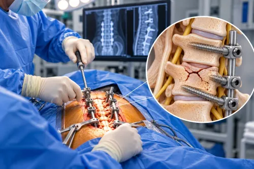
- Choose open incisional biopsy when the needle route is technically unsafe.
- Choose it after repeated nondiagnostic needle samples.
- Choose it when the sample cannot provide enough tissue for lymphoma, infection, or a suspected primary bone tumour.
- Avoid excisional biopsy for a possible primary malignant bone tumour unless an oncology surgeon has planned the operation; unplanned removal can compromise definitive surgery.
Preparation reduces preventable harm. Review anticoagulant and antiplatelet medicines with the procedural team and prescribing clinician; never stop them independently. Check the bleeding history, platelet count, coagulation results, allergies, pregnancy possibility, and any active infection before scheduling the procedure. Bleeding, infection, nerve injury, vascular injury, fracture, and a nondiagnostic sample remain possible risks.
A bone abnormality biopsy can fail if the needle enters necrotic tissue, reactive bone, a fracture cleft, or the adjacent disc instead of the enhancing solid component. Review the MRI and CT with the radiologist, target viable representative tissue, and plan enough cores for pathology, microbiology, flow cytometry, immunohistochemistry, or molecular testing.
These details often determine which spinal tissue biopsy choices remain useful.
Selecting the target and the specialist team
A biopsy should target the lesion that will answer the clinical question with the least risk, not automatically the most painful or largest abnormality. If several lesions exist, doctors compare MRI and CT for viable enhancing tissue, cortical destruction, accessibility and proximity to the cord, nerve roots and blood vessels.
A safer lesion outside the spine can sometimes establish the diagnosis; suspected primary spinal tumors require a tract planned for later excision.
| Target choice | Best use | Main safeguard |
|---|---|---|
| Most viable part of the spinal lesion | Suspected primary bone tumor or infection | Avoid necrosis, fracture clefts, reactive bone and adjacent disc |
| Safer accessible lesion elsewhere | Multiple lesions or suspected metastatic disease | Confirm that its result will change treatment |
| Cervical or technically difficult lesion | Lesion cannot be sampled safely from another site | Map arteries, airway, esophagus, cord and nerve roots on cross-sectional imaging |
Ask for a joint review before scheduling. The spine or orthopaedic oncologic surgeon plans the tract with an experienced musculoskeletal or interventional radiologist; a bone-tumor pathologist confirms how many cores and which ancillary tests are needed.
Send separate sterile specimens for bacterial, fungal and mycobacterial cultures when infection is possible, and request flow cytometry when lymphoma is a concern.
The surgeon who may perform definitive treatment must approve the route because a contaminated tract can complicate tumor removal or spine-sparing surgery. This coordination is central to deciding which spine biopsy approach is right for bone abnormality.
A spine biopsy specialist in Jaipur, such as Dr Rahul Temani, should help ensure imaging, sampling and treatment planning address the same lesion.
Related service
Advanced Spine Biopsy Treatment for Accurate Diagnosis Advanced Spine Biopsy Treatment for Accurate Diagnosis Spine Biopsy A spine biopsy is a minimally invasive diagnostic procedure used to collect a small sample of bone or tissue from the spine to detect... View service → |
Frequently asked questions
Does every vertebral lesion need a biopsy?
No. Doctors compare MRI and CT findings with symptoms, earlier scans, and risks such as cancer or infection. Biopsy becomes important when imaging cannot identify the lesion reliably or tissue results will change treatment.
What are the main spine biopsy options?
Fine-needle biopsy collects cells or fluid, core-needle biopsy removes a small cylinder of tissue, and open biopsy removes tissue through surgery. The choice depends on whether the laboratory needs cells, architecture, cultures, or a larger specimen.
How is the safest biopsy route through the spine chosen?
The team selects an entry path that reaches the target while avoiding the spinal cord, nerve roots, major blood vessels and vital organs. CT guidance helps plan and monitor many percutaneous procedures.
When is a percutaneous spine biopsy the wrong tool?
It may be unsuitable when the lesion cannot be reached safely, the sample would be too small, infection requires a larger specimen, or surgery is already needed for instability, compression or treatment.
Which specialists should select and perform a spine biopsy?
The plan should involve the clinician evaluating the lesion, an image-guided biopsy specialist or spine surgeon, a radiologist, and a pathology team. Ask how the specimen will be tested for tumour, infection and other diagnoses.
Keywords